
Western blotting is a powerful and widely used technique in molecular biology and biochemistry for detecting specific proteins in complex samples. Its mechanism involves a sequence of precise steps that enable researchers to separate, transfer, and identify proteins based on their size and antibody specificity. Understanding this mechanism is essential for interpreting experimental results and troubleshooting issues in protein analysis.
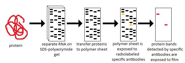
1. Protein Separation by Gel Electrophoresis
The first step in the mechanism of western blotting is the separation of proteins using gel electrophoresis. Typically, sodium dodecyl sulfate-polyacrylamide gel electrophoresis (SDS-PAGE) is used. SDS is an anionic detergent that denatures proteins and imparts a uniform negative charge proportional to the protein’s length. When an electric field is applied, western blot tutorials and examples migrate through the polyacrylamide gel matrix. Smaller proteins move faster and travel farther, while larger proteins migrate more slowly. This separation is crucial because it allows the identification of proteins based on molecular weight.
2. Transfer of Proteins onto a Membrane
Once proteins are separated, they are transferred from the gel onto a membrane, typically made of nitrocellulose or polyvinylidene difluoride (PVDF). This step is known as blotting. The transfer can be performed using either a wet transfer or semi-dry transfer system. An electric field drives the proteins out of the gel and onto the membrane, which immobilizes them in the same pattern as in the gel. This step is critical because the membrane provides a stable surface for protein detection, which is not possible directly in the gel.
3. Blocking Non-Specific Binding Sites
After transfer, the membrane contains exposed sites that can bind antibodies non-specifically. To prevent this, the membrane is incubated with a blocking solution, often containing proteins like bovine serum albumin (BSA) or non-fat dry milk. Blocking saturates these non-specific sites, ensuring that subsequent antibody binding is specific to the target protein. This step is essential to reduce background noise and improve the accuracy of detection.
4. Antibody Incubation
The core of the western blotting mechanism is the antibody-based detection of the target protein. The membrane is first incubated with a primary antibody that specifically binds to the protein of interest. After washing away unbound primary antibodies, a secondary antibody conjugated to an enzyme or fluorophore is applied. The secondary antibody binds to the primary antibody and facilitates visualization. This two-step antibody system enhances signal strength and specificity.
5. Detection and Visualization
Finally, the protein-antibody complexes are visualized using a detection method appropriate for the label on the secondary antibody. For enzyme-conjugated antibodies, chemiluminescent or colorimetric substrates are applied, producing light or a visible color where the target protein is located. Fluorescently labeled antibodies allow detection using imaging systems. The resulting image displays bands corresponding to the target protein’s molecular weight, providing qualitative and quantitative information about protein expression.
6. Data Analysis
After detection, densitometry or image analysis software can quantify the protein bands. This step allows researchers to compare protein levels across samples, study expression patterns, or evaluate post-translational modifications. Accurate data interpretation requires understanding the transfer efficiency, antibody specificity, and exposure conditions, all of which are integral to the western blotting mechanism.
Conclusion
The mechanism of western blotting integrates several biochemical principles, including protein separation, specific antibody-antigen recognition, and sensitive detection methods. By carefully following each step—from SDS-PAGE to visualization—researchers can reliably detect and quantify proteins in complex biological samples. Mastery of this mechanism not only facilitates protein research but also provides insights into cellular processes, disease mechanisms, and therapeutic targets.