
Introduction to Tissue Microarray
Tissue microarray (TMA) technology has revolutionized biomedical research and diagnostic pathology by enabling the simultaneous analysis of hundreds of tissue samples on a single slide. Since its introduction in the late 1990s, tissue microarray has become an essential tool in cancer research, biomarker discovery, drug development, and translational medicine. By allowing high-throughput analysis while conserving valuable tissue samples, TMA offers an efficient, cost-effective, and standardized approach to studying tissue-based molecular and histological changes.
In simple terms, a tissue microarray consists of tiny cylindrical cores extracted from multiple donor tissue blocks and arrayed into a single recipient paraffin block. Thin sections cut from this block can then be used for immunohistochemistry (IHC), in situ hybridization (ISH), fluorescence in situ hybridization (FISH), and other molecular techniques. This article explores tissue microarray technology in depth, including its methodology, advantages, applications, limitations, and future prospects.
What Is Tissue Microarray Technology?
Tissue microarray is a laboratory technique that allows researchers to analyze multiple tissue specimens in parallel under identical experimental conditions. Instead of processing each tissue sample individually, small tissue cores—typically ranging from 0.6 mm to 2.0 mm in diameter—are precisely extracted from donor blocks and re-embedded into a single paraffin block.
Each tissue core represents a specific patient, disease state, or experimental condition. When a section is cut from the tissue microarray block, it contains hundreds of different tissue samples arranged in a grid-like pattern. This structure enables rapid comparison and quantitative analysis across large cohorts.
History and Development of Tissue Microarray
The concept of tissue microarray was first introduced by Dr. Kononen and colleagues in 1998. Before the advent of TMA, tissue-based studies were labor-intensive, costly, and limited in scale. Researchers had to stain and analyze individual tissue slides, making large population studies impractical.
Tissue microarray technology addressed these challenges by combining miniaturization with automation. Over time, advancements in arrayers, imaging systems, and digital pathology software have significantly improved the accuracy, reproducibility, and analytical power of TMAs. Today, tissue microarray is a standard method in pathology labs worldwide.
How Tissue Microarray Is Constructed
Selection of Donor Tissue
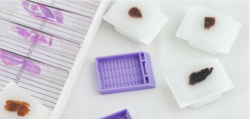
The first step in tissue microarray construction involves selecting appropriate donor tissue blocks. These blocks are usually formalin-fixed, paraffin-embedded (FFPE) samples obtained from biopsies or surgical resections. A pathologist reviews hematoxylin and eosin (H&E)-stained slides to identify representative areas of interest, such as tumor regions or normal tissue controls.
Core Extraction and Arraying
Using a tissue microarrayer (manual or automated), small cylindrical cores are punched from the selected regions of donor blocks. These cores are then precisely inserted into pre-defined coordinates in a recipient paraffin block. Each position is mapped and documented to ensure accurate sample identification.
Sectioning and Staining
Once the tissue microarray block is completed, thin sections (typically 3–5 microns thick) are cut using a microtome and mounted onto glass slides. These slides can be stored or immediately processed for various staining and molecular analysis techniques.
Applications of Tissue Microarray
Cancer Research
Cancer research is the most prominent application of tissue microarray technology. TMAs allow researchers to analyze protein expression, gene amplification, and molecular alterations across hundreds of tumor samples simultaneously. This capability is critical for identifying diagnostic, prognostic, and predictive biomarkers.
For example, tissue microarray is widely used to study biomarkers such as HER2 in breast cancer, p53 mutations, Ki-67 proliferation index, and hormone receptor status. Large-scale studies using TMAs help correlate molecular findings with clinical outcomes, leading to more personalized treatment strategies.
Biomarker Discovery and Validation
Tissue microarray plays a vital role in biomarker discovery and validation. Once a potential biomarker is identified, TMAs enable rapid screening across large patient cohorts to confirm its clinical relevance. Because all tissue samples are processed under identical conditions, variability is minimized, improving data reliability.
Drug Development and Pharmacological Studies
Pharmaceutical companies and academic researchers use tissue microarray to evaluate drug targets, mechanisms of action, and treatment response. TMAs help assess how different tissues or tumor subtypes respond to experimental therapies, accelerating preclinical and translational research.
Diagnostic Pathology
In diagnostic pathology, tissue microarray serves as a valuable quality control and educational tool. Pathologists use TMAs to standardize staining protocols, compare antibody performance, and train residents by presenting multiple disease entities on a single slide.
Genomic and Proteomic Research
Tissue microarray integrates seamlessly with genomic and proteomic technologies. Techniques such as FISH, chromogenic in situ hybridization (CISH), and RNA-based assays can be applied to TMA sections, allowing researchers to study gene expression and chromosomal alterations in situ.
Advantages of Tissue Microarray
High-Throughput Analysis
One of the greatest advantages of tissue microarray is its high-throughput capability. Hundreds of tissue samples can be analyzed simultaneously, dramatically reducing time and labor compared to conventional methods.
Cost Efficiency
By consolidating multiple samples onto a single slide, tissue microarray significantly reduces reagent consumption, antibody usage, and processing costs. This makes large-scale studies more economically feasible.
Standardization and Reproducibility
Since all samples on a tissue microarray slide undergo identical processing and staining conditions, experimental variability is minimized. This standardization improves reproducibility and comparability of results.
Conservation of Valuable Tissue
Clinical tissue samples are often limited and irreplaceable. Tissue microarray conserves precious tissue by using only small cores, allowing donor blocks to be preserved for future studies.
Limitations of Tissue Microarray
Despite its many advantages, tissue microarray technology has certain limitations that researchers must consider.
Sampling Bias
Because TMAs analyze only small tissue cores, they may not fully represent the heterogeneity of a tumor or tissue. This is particularly relevant for cancers with marked spatial variability. Using multiple cores per case can help mitigate this issue.
Technical Challenges
Accurate construction of tissue microarrays requires expertise and precision. Core loss, misalignment, and sectioning artifacts can occur if proper techniques are not followed.
Limited Use for Rare Structures
Some rare or focal pathological features may be missed in tissue microarray cores, making whole-section analysis more appropriate in certain cases.
Tissue Microarray and Digital Pathology
The integration of tissue microarray with digital pathology has further expanded its utility. High-resolution slide scanners and image analysis software enable automated quantification of staining intensity, cell counts, and spatial distribution of biomarkers.
Artificial intelligence (AI) and machine learning tools are increasingly being applied to tissue microarray datasets, allowing more objective and scalable data analysis. This combination is transforming pathology from a subjective, manual discipline into a data-driven science.
Ethical and Regulatory Considerations
The use of human tissue in tissue microarray research requires adherence to ethical and regulatory guidelines. Informed consent, institutional review board (IRB) approval, and data anonymization are essential components of responsible TMA research. Proper documentation ensures patient privacy while enabling valuable scientific insights.
Future Directions of Tissue Microarray Technology
The future of tissue microarray looks promising, with ongoing innovations aimed at improving resolution, automation, and integration with multi-omics platforms. Emerging developments include:
- High-density tissue microarrays with thousands of cores per block
- Multiplex staining techniques for simultaneous detection of multiple biomarkers
- Integration with spatial transcriptomics
- AI-powered image analysis for predictive modeling
As precision medicine continues to evolve, tissue tissue samples will remain a cornerstone technology for linking molecular data with clinical outcomes.
Conclusion
Tissue microarray technology has fundamentally transformed tissue-based research by enabling high-throughput, cost-effective, and standardized analysis of large sample cohorts. From cancer research and biomarker discovery to diagnostic pathology and drug development, tissue microarray continues to play a critical role in advancing medical science.
While it has certain limitations, careful experimental design and technological advancements have significantly enhanced its reliability and applicability. As digital pathology and artificial intelligence converge with tissue microarray methods, this powerful technology will continue to drive innovation in personalized medicine and translational research.